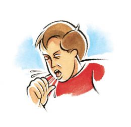
Common Recurrent or Persistent Respiratory Symptoms –
- Cough
- Wheeze
- Stridor
- Persistent Lung Infiltrates #
Evaluation of chronic respiratory symptoms can be difficult because –
- Symptoms – close succession of unrelated ARTI
- Symptoms may have Single pathophysiologic process.
- Lack of specific diagnostic tests.
- Parental anxiety – pressure for quick therapeutic relief.
Approach to Diagnosis and Treatment
•Points to remember
–Symptoms – minor problem or life threatening process.
–Determine – most likely pathogenic mechanism.
–Simplest effective therapy for the underlying process- often may be symptomatic only.
–Occasionally , use of more extensive and invasive diagnostic efforts including bronchoscopy.–Evaluation of the effect of therapy.
When Chronic Respiratory Complaints Can be Life Threatening/ Serious?
- Persistent fever
- Ongoing limitation of activity
- Failure to grow
- Failure to gain weight appropriately
- Clubbing of the digits
- Persistent tachypnea and labored ventilation
- Chronic purulent sputum
- Persistent hyperinflation
- Substantial and sustained hypoventilation

- Refractory infiltrates on CXR
- Persistent pulmonary function abnormalities
- Family h/o heritable lung disease
- Cyanosis and hypercarbia
Causes of Recurrent or persistent Wheeze
- Wheeze associated lower respiratory tract infection.
- Bronchiolitis and Bronchial asthma
- Tropical eosinophilia
- Loeffler’s syndrome
- Inhaled foreign bodies
- Hypersensitivity pneumonitis
- External compression
- Cystic fibrosis
Causes of Stridor in children
•Recurrent
–Allergic croup ( Spasmodic Croup)
–Laryngomalacia
–Respiratory infections in a child with otherwise asymptomatic anatomic narrowing of the large airways
•Persistent
–Laryngeal obstruction- laryngomalacia, laryngeal webs, cysts and laryngoceles, foreign body.
–Tracheobronchial disease- tracheomalacia, subglottic tracheal webs & stenosis, extrinsic masses
•Others
–Gastroesophageal reflux, Pierre Robin syndrome, macroglossia, hypocalcemia, hysterical stridor
Evaluation of Stridor
Physical examination -unrewarding, although changes in its severity and intensity due to changes of body position should be assessed.
Anteroposterior and lateral roentgenograms
In most cases, direct observation by laryngoscopy is necessary for diagnosis. Undistorted views of the larynx are best obtained with fiberoptic laryngoscopy.
Others-Contrast esophagography,Fluoroscopy, CT, and MRI are potentially useful diagnostic tools.
COUGH
Cough is one of the most common symptoms for which outpatient care is asked, accounting for up to 40% of the practice activity. [Irwin RS Et Al Chronic cough. The spectrum and frequency of causes, key components of the diagnostic evaluation, and outcome of specific therapy. Am Rev Respir Dis 1990, 141:640–647.]
Cough is a reflex response of the lower respiratory tract to stimulation of irritant or cough receptors in the airways’ mucosa.
Receptors- pharynx, paranasal sinuses, stomach, and external auditory canal, the source of a persistent cough may need to be sought beyond the lungs.
Stimuli include excessive secretions, aspirated foreign material, inhaled dust particles or noxious gases, and an inflammatory response to infectious agents or allergic processes
Cough Reflex
Inspiratory phase: Inhalation generates the volume necessary for an effective cough.
Compression phase: Closure of the larynx combined with contraction of muscles of chest wall, diaphragm, and abdominal wall result in a rapid rise in IT pressure.
Expiratory phase: The glottis opens, resulting in high expiratory airflow and the coughing sound.
The high flows dislodge mucus from the airways and allow removal from the tracheobronchial tree
Causes of recurrent or persistent cough
Very common – Asthma, Recurrent infections.
Common – Prolonged infection, cigarette smoking, habit cough, Post-infective (adenovirus, pertussis), Tuberculosis
Uncommon – Aspiration ( GERD, Uncoordinated swallowing), Intrabronchial/ retained foreign body, Cystic fibrosis, ciliary abnormalities, immunodeficiency , Congenital abnormalities of respiratory tract.
Rare – Mediastinal or Pulmonary tumors
Evaluation- History
A detailed history should include following questions
•How and when did the cough start?
•What is the nature and quality of the cough?
•Is the cough an isolated symptom?
•What triggers the cough?
•Is there a family history of respiratory symptoms, disorders and atopy?
•What medications is the child on?
•Does the cough disappear when asleep?
•Does the child smoke cigarettes or exposed to environmental smoke? (BTS Guidelines)
Age- in relation to cough
| Age at Onset |
Likely causes |
| Onset from birth |
Laryngeal web, vascular ring, H type TEF |
| Starting in 1st month |
congenital infections( Rubella,CMV) leading to interstitial pneumonia |
| In Early infancy- |
GERD, Recurrent Aspirations |
| In Late infancy- |
Bronchiolitis, Asthma, cystic fibrosis, Pertusis |
| In preschool age- |
Recurrent bronchitis, allergic bronchitis, asthma, foreign body, chronic suppurative lung disease, pulmonary eosinophilia |
| At all ages- |
Asthma, whooping cough, viral bronchitis, tuberculosis, foreign body aspiration |
Duration of Symptom: Classification
Acute if < 3 weeks duration Duration- chronic if >3 wk
Chronic Cough- defined as a cough of >4 weeks [Guidelines for Evaluating Chronic Cough in Pediatrics ACCP Evidence-Based Clinical Practice Guidelines 2006]
Acute cough- A recent onset of cough lasting <3 weeks.
Prolonged acute cough- resolving over a 3–8-week period.
Chronic cough – A cough lasting >8 weeks.
Recurrent cough- repeated (>2/year) cough episodes, apart from those associated with colds, each lasting more than 7–14 days. [Recommendations for the assessment and management of cough in children, British Thoracic Society Cough Guideline Group 2007]
Onset –
Acute and persisted- Retained Inhaled FB
Started with cold – Infective
Character of Cough
| Cough character |
Diseases |
| Staccato, paroxysmal |
Pertussis, cystic fibrosis, foreign body, Chlamydia spp., Mycoplasma spp. Followed by “whoop” Pertussis |
| All day, never during sleep- |
Habit (tic) cough |
| Barking, brassy – |
Croup, psychogenic, tracheomalacia, tracheitis, epiglottitis |
| Hoarseness |
Laryngeal involvement (croup, recurrent laryngeal nerve involvement) |
| Abrupt onset |
Foreign body, pulmonary embolism Follows exercise Reactive airways disease |
| Accompanies eating, drinking |
Aspiration, gastroesophageal reflux, tracheoesophageal fistula |
| Throat clearing |
Postnasal drip, vocal tic |
| Productive (sputum) |
Infection |
| Night cough |
Sinusitis, reactive airways disease ,Seasonal Allergic rhinitis. |
| Immunosuppressed patient |
Bacterial pneumonia, Pneumocystis jiroveci, PTB, MAC, CMV |
| Animal exposure |
Chlamydia psittaci (birds), Francisella tularensis (rabbits), histoplasmosis (pigeons) |
| Geographic |
Histoplasmosis (Mississippi, Missouri, Ohio River Valley), coccidioidomycosis (southwest), blastomycosis (north and midwest) |
| Workdays with clearing on days off |
Occupational exposure |
Progression-
Relentlessly progressive- Inhaled foreign body, Lobar collapse, Tuberculosis, Rapidly expanding intrathoracic lesion
Triggering factors-
•Exercise, cold air, early morningà Asthma
•Lying downà Postnasal drip, gastro-oesophageal reflux disease
•Feeding à Recurrent pulmonary aspiration
Associated History
Associated with difficulty in breathing or not
Sputum- mucoid, mucopurulent, thick, tenacious or viscous, hemoptysis ( Pneumonia, lung abscess, bronchitis,bronchiectasis)
Chest pain- Asthma, functional, pleuritis
Fever- Infections
Recent Weight loss- lung abscess, PTB
Effect of season- Allergic etiology
Preceding event- ingestion of kerosene, choking episodes
About symptoms of Tuberculosis, ENT diseases
Cardiac diseases *
Failure to thrive- Compromised lung function, immunodeficiency cystic fibrosis
Past history – recurrent rhinitis, atopic dermatitis, eczema, pertussis, recurrent or severe respiratory illness in infancy or childhood (Immunodeficiency, CF, bronchiectasis).
Birth history – Prematurity, asphyxia, neonatal intubation, prolonged ventilatory support, delayed meconium stool. Maternal history of abortion, rashes, lymphadenopathy during pregnancy
Immunization history ( Hib, pneomococcal vaccine) including Vitamin A supplementation
Feeding history – Exclusive breast feeding, Association of symptom with weaning, Cows milk
Delayed milestones of development
Treatment history – Enalapril (bradykinin cough), allergy
Family history – size, overcrowding, respiratory disease, atopy
Domestic smoke pollution including parental smoking.
Personal history- smoking,alcohol
Exposure to allergens – pets
H/o eating crabs – Paragonimiasis
Deworming
H/O consanguinity
CLINICAL EXAMINATION
1.General appearance:-
Consciousness and orientation
Tachypnea, use of accessory muscle – respiratory distress.
2. Vital signs :-
Fever – infective pathology.
Pulse – tachycardia–CCF, fever, distress.
SpO2- hypoxia
3. Other Signs-
Cyanosis – hypoxemia/heart disease.
Digital clubbing- Suppurative lung disease, CF, Bronchiectasis
Edema- cardiac disease
JVP – raised in CCF.
4. Anthropometry :Evidence of FTT – consider CF, immunodeficiency.
Subconjunctival hemorrhage – pertussis
5. ENT Examination:-
Nose – nasal discharge – blood stained/ serous/ purulent; DNS.
Polyps – allergy.
Pharynx – congested/ grey membrane.
Tonsils – enlargement/ congestion/pus points.
Ear – discharge (ASOM), retraction of TM.
6. Signs of atopic disease :-
•Eczema, transverse nasal crease, rhinitis, mucosal cobblestoning, injected conjunctivae – consider RAD, allergy.
7. Sinusitis:-
•Periorbital edema, sinus tenderness, purulent posterior pharyngeal drainage, halitosis
Systemic Examination-
Respiratory system :-
Inspection-
•Intercostal or subcostal indrawing.
•Intercostal fullness or crowding of ribs
•Decreased movement of either hemithorax.
•Suprasternal recessions – suggestive of narrowing or obstruction of upper airways
•Position of trachea
Grunting, nasal flaring, head nodding, wheezing
Stridor, in combination with cough, generally indicates obstruction at the level of larynx or trachea.
Palpation
•Feel for abnormal vibrations – rhonchi, friction rub, crackles, crepitus (subcutaneous emphysema- pertussis).
•Vocal fremitus.
•Expansion of hemithorax.
Percussion
•Area of dullness- pleural effusion , empyema, consolidation.
•Hyperresonant – pneumothorax.
•Percuss for upper margin of liver dullness.
Auscultation
•Compare air entry B/L.
•Bronchial breath sounds.
•Added sounds – crackles, rhonchi, pleural friction rub.
•Vocal resonance
-absent/ decreased – pleural effusion, atelectasis.
-Increased – consolidation, atelectasis with patent bronchus.
Cardiovascular System-
–Apex beat
–Palpable P2
–Thrill, Heave,Murmur
–S3,S4
Abdomen
–Palpable liver- Hyperinflated chest or CCF ( tender hepatomegaly)
CNS, Musculoskeletal system
Assessment
•Specific cough- A specific cough is one in which there is a clearly identifiable cause.
•Non-specific (isolated cough) – The term ‘‘non-specific isolated cough’’ has been used to describe children who typically have a persistent dry cough, no other respiratory symptoms (isolated cough), are otherwise well with no signs of chronic lung disease and have a normal chest radiograph.
INVESTIGATIONS
•Chest Radiograph
•HRCT of the Chest
•Pulmonary Function Tests
•Gastrointestinal Studies
•Sinus Imaging
•Laryngoscopy
•Bronchoprovocation Test
•Bronchoscopy
CHEST RADIOGRAPH
•A chest radiograph is indicated for most children with chronic cough.
–overview of the state of the lungs
–normal chest radiograph does not always exclude significant pathology such as bronchiectasis
–further lab studies.
–Also helpful in identifying extra pulmonary causes.
Normal X-ray: Asthma, GERD, Postnasal drip, Bronchitis and upper airways disease.
•Not indicated if mild specific disorder is definitively diagnosed (asthma/allergic rhinitis or if a pertussis-like illness is clearly resolving).
HRCT-CHEST
•May identify parenchymal disease not apparent on chest X-ray.
•Useful in the diagnosis of disorders of smaller airways e.g. bronchiectasis (Gold standard)-more sensitive than spirometric indexes.
•Sinus CT scans have definite roles- Uncommonly prescribed
•Balance- Benefit vs Hazard
PULMONARY FUNCTION TESTS
•Includes peak flow meter & spirometric examination.
•Useful in evaluation of children with chronic cough and normal chest X-ray.
•Peak expiratory flow (PEF) measurement should be focused on the diagnosis of airflow obstruction.
•PEF measurement to assess bronchodilator response have limitations compared to FEV1.
Age- children aged 6 years and in some children 3 years if trained pediatric personnel are present.
Spirometry-
–FEV1/FVC < 0.8 (airflow obstruction)
–Improvement in FEV1 after bronchodilator therapy by > 15%*
–Exercise challenge – worsening in FEV1 >15% *
PEF morning to evening variation >20%*
Also detects – restrictive lung diseases
*- main criteria consistent with asthma
GASTROINTESTINAL STUDIES
•24- Hour esophageal pH monitoring- high sensitivity & specificity.
•Negative pH profile reports should be interpreted cautiously.
•Dual probe pH monitoring has improved the sensitivity of esophageal pH monitoring in patients who have non- acid reflux.
•Barium swallow- low sensitivity & specificity.
LARYNGOSCOPY
•Both direct & fiberoptic – laryngoscopy very useful.
•A thorough ENT examination & laryngoscopic examination complements each other in diagnosing upper airway causes of persistent cough.
BRONCHOPROVOCATION TEST
•Hypersensitivity to methacholine is useful in evaluating patients with possibility of cough variant asthma.
•Although a positive test is not diagnostic of asthma, a negative test effectively excludes asthma as the diagnosis.
BRONCHOSCOPY
•Flexible bronchoscope- evaluating upper and lower airway structure and function.
•Useful in identifying unrecognized FB in bronchus and airway sampling either by mucosal biopsy or bronchoalveolar lavage.
•Bronchoalveolar lavage useful in identifying organisms not found on culturing of sputum and blood and unusual organisms resulting in parenchymal disease.
•Both can yield useful information that can modify the treatment.
•Fibre- optic bronchoscopy- 64% sensitivity in children.
TREATMENT
•Effective management is possible by establishing a precise diagnosis.
•Systematic approach- effective in identifying an underlying cause in > 80 % cases.
•Establish diagnosis & institute appropriate specific therapies.
•If the putative specific therapy does not stop the cough, the presumptive diagnosis is either wrong or therapy submaximal.
SPECIFIC THERAPY
•Asthma/ cough variant asthma – bronchodilator therapy, inhaled corticosteroids, other asthma therapies.
•GERD – positional therapy, dietary modifications, anti-reflux therapy, surgical correction.
•Sinusitis – antibiotics, decongestive therapy, intranasal decongestive/ steroid spray.
•Bronchiectasis – chest physiotherapy/ postural drainage, judicious use of antibiotics, expectorants/ mucolytics, bronchodilator therapy, surgical resection.
•Psychogenic & habitual cough – counseling and psychological therapy, avoidance of mental stress.
•Lung abscess – judicious use of antibiotics, removal of precipitating cause, surgical intervention if necessary.
•TB – ATT.
•Foreign body aspiration – FB removal, antibiotics, physiotherapy and exercises.
•Post acute viral respiratory infection – symptomatic treatment, bronchodilators, oral steroid therapy.
•Pertusis – macrolides,symptomatic treatment.
•Congenital anomalies/ malformations – surgical corrections, symptomatic & supportive therapy.
•Drug induced cough – stop the drug.
Recurrent and Persistent Lung Infiltrates
•Radiographic lung infiltrates that fail to completely clear within a 4-wk period.
•May be febrile or afebrile and may display a wide range of respiratory symptoms and signs.
•Persistent or recurring infiltrates present a diagnostic challenge
•Symptoms associated with chronic lung infiltrates in the 1st several weeks of life (but not related to neonatal respiratory distress syndrome) à Congenital infections
•Early appearance of chronic infiltratesàCF, congenital anomalies, aspirations
•Recurrent infiltrates, wheezing, and coughà asthma, even in the 1st year of life.
•1st year of life with recurrent lung infiltratesà pulmonary hemosiderosis .
•Recurrent pneumonia,frequent otitis media, nasopharyngitis, adenitis, or dermatologic manifestations àimmunodeficiency state LIP in HIV
•Paroxysmal coughing à pertussis syndrome or CF.
Persistent infiltrates, especially with loss of volume, in a toddler à foreign body aspiration.
•A “silent chest” with infiltrates à alveolar proteinosis , Pneumocystis jiroveci infection
•Cataracts, retinopathy, or microcephaly suggest in utero infection.
Diagnostic studies as suggested by history and physical examination
•Cytologic evaluation of sputum
•Chest CT -precise anatomic detail concerning the infiltrate.
•Bronchoscopy is indicated for detecting foreign bodies, congenital or acquired anomalies of the tracheobronchial tract, and obstruction by endobronchial or extrinsic masses.
•Optimal medical or surgical treatment of chronic lung infiltrates often depends on a specific diagnosis
•symptomatic therapy – post viral
•inhalation and physical therapy for excessive secretions, antibiotics for bacterial infections, and maintenance of adequate nutrition.
PROGNOSIS
•Dependent on etiology.
•An appropriate diagnosis and effective specific therapy- associated with good prognosis with cure rate of 84-97%.
•Prognosis good in single etiology cases than multiple etiology cases.
•A systemic approach both for the diagnosis and treatment- essential for better prognosis.
•Poor prognosis- misdiagnosis & inappropriate therapy.
REFERENCES
•Behrman,Kliegman & Jenson, NelsonTextbook Of Pediatrics ,19th edition.
•Ghai, O P ; Essential Pediatrics ,7th edition.
•Guidelines for Evaluating Chronic Cough in Pediatrics ACCP Evidence-Based Clinical Practice Guidelines
•Recommendations for the assessment and management of cough in children M D ShieldsEt al British Thoracic Society Cough Guideline Group
•Perspective on the human cough reflex-Stuart M Brooks, COUGH 2011
•Bronchoscopic Findings in Children With Chronic Wet Cough, Pediatrics 2012


