Microbiology of Vibrio Cholera
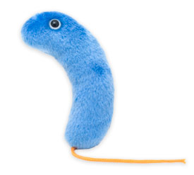 Vibrio cholerae (Kommabacillus) is the causative agent of Cholera. Cholera is an acute diarrhoeal infection caused by ingestion of food or water contaminated with the bacterium Vibrio cholerae. Every year, there are an estimated 3–5 million cholera cases and 100000–120000 deaths due to cholera. The appelation cholera probably derives from the Greek word for the gutter of a roof, comparing the deluge of water following a rainstorm to that from the anus of an infected person.
Morphology:
- Gram negative, non-sporing, non-capsulated, curved or comma-shaped rods
- Highly motile due to single polar flagellum and the movement is of a “darting type”
- In liquid cultures they may be arranged singly, in pairs or in S-shaped spiral forms
- Pleomorphism is common in old cultures
- 2 X 0.5 micrometer in size
Classification:
A) V.cholerae O1:
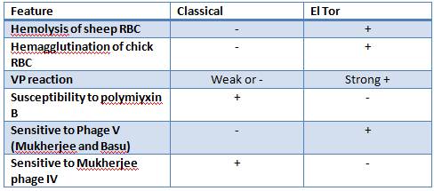
- Classical and El Tor biotypes
Each biotype can be further divided into Ogawa, Inaba and Hikojima strain. The strains and the difference in their O antigens (A,B,C) are:
- Ogawa: AB
- Inaba: AC
- Hikojima: ABC
B) Non-O1 V.cholerae group:
Until recently only the O1 serotype was associated with severe diarrhea. Beginning in the 1992, a new V.cholerae serotype (O139, also known as Bengal) has been associated with severe, watery diarrhea.
Pathogenesis:
The vibrios never invade the epithelium but instead remain within the lumen and secrete an enterotoxin which is encoded by a virulence phage.
A) Entry into body:
To establish disease, V. cholerae must be ingested in contaminated food or water and survive passage through the gastric barrier of the stomach.
B) Bacterial Colonization:
- Fllagelar proteins mediate chemotaxis based motility which helps virulent V.cholerae to reach the luminal epithelium in small intestine
- Proteolytic enzymes help to dissolve glycoprotein coating of the intestinal cells.
- Coregulated pili along with accesory colonization factor “adhesin” mediate adherence of the organism to the epithelium of microvilli at the brush border where they multiply.
- Hemagglutination-protease (mucinase) is important for detachment of Vibrio from epithelial cells
C) Secretion of cholera toxin:
- The choleragen enterotoxin is released in close proximity to its specific receptor (GM1 ganglioside) on cell membrane lining the villi and crypts of small intestine.
- Cholera toxin is composed of 5 binding subunits (B) and 1 active catalytic subunit (A).
D) Activation of the A1 subunit: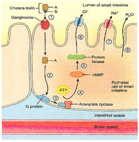
- The B subunits, serving as “landing pad”, bind to carbohydrates on GM1 ganglioside on the surface of the eopithelial cells of the small intestine, enabline caveolar-mediaed endosomal entry of toxin subunit A into the cell.
- The reverse transport of subunit A from endosome into the cell cytoplasm is followed by cleavage of the disulfide bond linking the 2 fragments of peptide A (A1 and A2).
- The generated catalytic peptide A1 forms complex with ADP ribosylation factors (ARF) which catalyzes the ADP ribosylation of a G protein.
- Binding of NAD and GTP generates an activated G protein which in turns bind to and stimulates adenylate cyclase.
- The activated adenylate cyclase generates high levels of intracellular cAMP from ATP.
- cAMP (Cyclic AMP) stimulates secretion of chloride and bicarbonate, with associated sodium and water secretion. Chloride and sodium resorption are also inhibited.
E) Result:
- “Rice water” diarrhea containing flecks of mucus – upto 14 L/day, equivalent to the circulating blood volume causes severe dehydration and electrolyte imbalances.
Clinical syndrome:
- Occurs 2-3 days after consumption of contaminated food/water
- Ranges from asymptomatic to the most severe form, known as “cholera gravis”
- 75% asymptomatic
- 20% mild disease
- 2-5% severe
- Clinical features include:
- Vomiting
- Cramps
- Sunken eyes
- Profuse “rice-water” stools (1L/hr) followed by rapid dehydration and hypovolemic shock
- No fever since the bacteria is non-invasive
- Without treatment, death occurs within 18 hours to several days
Consequences of severe dehydration:
- Intravascular volume depletion
- Severe metabolic acidosis
- Hypokalemia
- Cardiac and renal failure
Epidemiology:
- Fecal-oral route route of transmission and principally water-borne
- Endemic in areas of poor sanitation (India, Sub-Saharan Africa, Southern Asia)
- May persist in shellfish or plankton
- 7 pandemics since 1817: First 6 from Classical biotype and 7th from the El Tor biotype
- In 1993 Cholera epidemic in Bengal was caused by O139 which may be the cause of the 8th pandemic
- The recent Cholera epidemic in Haiti occured due to El Tor O1 strains that are predominant in South Asia.
Laboratory Diagnosis:
A) Specimen:
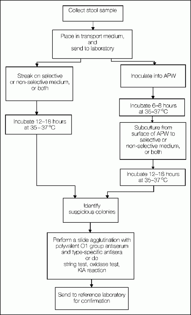
- Watery stool and mucus flakes from stool
- Rectal swab
- Vomitus
- Strip of blotting paper soaked in watery stool
B) Preservation and Transport media:
When there may be delay in transmission of specimen to laboratory, it must be refrigerated at 8c to 10c for 24 hours, else following transport media may be used:
- Vekataraman-Ramakrishnan (V.R) medium
- Cary-Blair medium
Subcultures are made from transport media to TCBS or BSA within 12-18 hours since other organisms may begin to overgrow the media after prolonged incubation.
C) Enrichment media:
- Alkaline peptone water (pH 8.6)
- Monsur’s taurocholate tellurite peptone water (pH 9.2)
D) Microscopy:
Direct microscopy: Hanging drop preparation to see the motility of the bacteria
Gram staining: Gram-negative, short-curved (comma-shaped) rods
Fluorescent antibody staining: for identifying V.cholerae O1
E) Culture:
Direct plating in selective media:
- Thiosulphate citrate bile sucrose agar (TCBS) aka Indicator-bromothymol blue medium: After overnight incubation at 37c, the colonies are large, moist, translucent, round and yellow due to sucrose fermentation and turns green on continuous incubation
- Alkaline Bile salt agar (BSA): colonies resembling that in the nutrient agar
- Monsur’s Gelatin Taurocholate Trypticate tellurite Agar (GTTA): small translucent colonies with a greyish black center.
Indirect plating:
The specimens received on transport medium are first inoculated into enrichment media and incubated for 6-8 hours before plating on selective medium.
F) Identification:
1) Biochemical tests:
- Ferments sucrose, glucose and mannose with production of acid and gas but fails to ferment arabinose
- Catalase and oxidase positive
- Cholera red reaction: When vibrio cholerae are grown for 24 hours in peptone water medium containing adequate amount of tryptophan and nitrate, they produce indole and reduce nitrate to nitrite. On adding a few drops of sulphuric acid, nitroso-indole is formed, which is red in color.
- Voges-Proskauer (VP) reaction: positive in El Tor biotype but negative in Classical biotype
2) Hemolysis reaction: The classical biotype do not hemolyse sheep or goat RBCs but El Tor biotype can hemolyse it.
3) Agglutination test:
Slide or Tube agglutination test with V.cholerae O1 antiserum:
- Positive test result: Test is repeated using monspecific Ogawa and Inaba sera. Hikojima strain react equally well with both Ogawa and Inaba sera.
- Negative test result: Tested with other O antiserum to establish their identity (non O1 strains)


2 Comments
SEND ME PDF ABOUT E.COLI SHIGELLA AND SALMONELLA AND VIBRIO CHOLARE BACTRIAS MORPHOLOGY ,CULTURAL PROPERTY , AND ETIOLOGY AND BIOCHEMICAL TESTS AND PATHOGENESIS
Hello. I would like to ask as question. which Agar is best for culturing Vibro cholerae in a medical laboratory?
Comments are closed.