Microbiology of Dermatophytes
Dermatophytosis:
Tinea or ringworm (dermatophytoses) are defined as cutaneous fungal infections of keratinized tissues (hair, nail, skin,etc.) by a group of related mould fungi called dermatophytes. Skin acts as a barrier to fungal infection. The fatty acid content and acid pH of skin, ciliary mucosal epithelia of respiratory tract and humoral factors restrict the growth of dermatophytes to the outer layers of skin.
Common dermatophytes and their features:
| Features | Trichophytom | Microsporum | Epidermophyton |
| Site of infection | Skin, hair, nail | Skin, hair | Skin, nail |
| Macroconidia | Sparse, thin-walled, smooth, Pencil shaped | Numerous, thick-walled, rough, Spindle shaped | Numerous, smooth-walled, Club shaped |
| Microconidia | Abundant | Rare | Absent |
| Examples | T.rubrum, T.schoenleinii, T.violaceum, T.concentricum, T.mentagrophytes, T.tonsurans, T.equinum | M.canis, M.fulvum, M.nanum, M.andouinii, M.gypseum, M.racemosum, M.equinum, M.gallinae | E.floccosum |
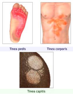
Clinical Forms:
Dermatophytes grow only on the keratinized layers of epidermis and their appendages. Usually they do not invade living tissues. These fungi secrete an enzyme called keratinase, which digests keratin. Since keratin is the primary structural protein of skin, nails, and hair, the digestion of keratin manifests as scaling of the skin, loss of hair, and crumbling of the nails.

| Dermatomycosis | Site Involved | Lesions | Causative Agent |
| Tinea capitis | Scalp | Scaly red lesions with loss of hair | Trichophyton, Microsporum (most species)
T.schoenleinii causes favus |
| Tinea corporis | Non-hairy skin of the body | Ring shape with red raised border | T.rubrum |
| Tinea cruris (Jock itch) | Groin and perineum | Itchy red patches | E.floccosum |
| Tinea barbae | Bearded areas of face and neck | Kerion like plaques | Trichophyton (most common) |
| Tinea pedis | Sole and toe clefts (Athlete’s foot) | Cracking and peeling of the skin | T.rubrum |
| Tinea unguim (Onychomycosis) | Nails | Thickened, discolored and brittle nails | Trichophytom (many species) and Epidermotophyton |
Laboratory Diagnosis:
Specimen: Nail samples, scrapings from the edges of skin lesions, plucked infected hairs
Direct Microscopy:
- Keratinized specimen is placed on a slide
- A drop of 10 to 20% KOH is added to dissolve keratin and cellular material, leaving the alkali resistant fungi intact
- Covered by a cover slip
- Left for 20 minutes in incubator at 37 c to digest keratin
- Microscopy reveals filamentous branching hyphae and arthrospore is oftenly seen in hair
Culture:
The specimen is inoculated into a slide or in a plate of Sabourad’s dextrose Agar (SDA) containing chloramphenicol or cycloheximide. Slightly acidic nature of SDA allows pathogenic fungi to grow but doesn’t allow bacterial growth. And antibiotics in the culture medium further prevent bacterial or saprophytic fungal contamination.
The slide/plate is incubated aerobically at 25-30c upto 3 weeks
Identification:

Trichophyton:
- Colonies show colors
- Examination of macro and microconidia
Microsporum:
- Microsporum on scalp can be identified because of bright green appearance of infected hair with an ultraviolet Wood’s light
- Examination of macro and microconidia
Epidermophyton:
- Colonies may appear white, yellow or olive in color
- Examination of macro and microconidia
Treatment:
- Topical imidazoles as 1st line drugs
- Oral griseofulvin for tinea capitis and tinea unguium