
How to Read a Chest Xray II : Pneumonia
Before Proceeding to How to Read Chest Xray of Pneumonia patient , read the sequential reading of chest Xray.
Terminologies:
RUZ- Right Upper Zone
RMZ- Right Middle Zone
RLZ- Right Lower Zone
CP angle- Costophrenic Angle
Air Bronchogram- air-filled bronchi (dark) being made visible by the opacification of surrounding alveoli (grey/white)
1. Pneumonia Lobar consolidation – usually Streptococcal pathology
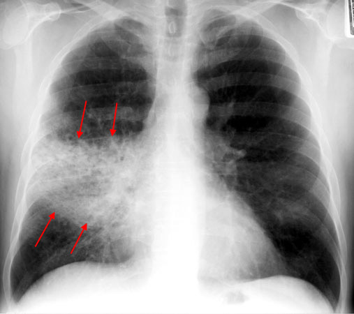
These are PA film of RMZ pneumonia.
Features of Pneumonia are-
- Normal or Increased Volume
- No Shift of Trachea
- Consolidation, Air Space Process
- Not Centered at Hilum
While Reading it Start sequentially . Here the findings can be read as-
To the Sequence add the finding ” The Trachea is Central, There is no shifting of Mediastinum, The Costophrenic angles are sharp and clear. There is non-homogenous opacity involving right middle zone with visible air bronchograms and indistinct borders. There is silhouetting of Right cardiac border ( RML pneumonia). Cardiac size looks Normal.”
2. Interstitial pneumonia : Usually Viral, Atypical organisms
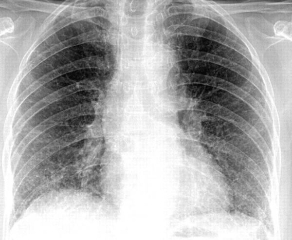
Involvement of the supporting tissue of the lung parenchyma resulting in fine or coarse reticular opacities or small nodules.
Findings ” Central trachea and Mediastinum, Diffuse fine reticular opacities involving the entire lung field with Normal CP angles and Normal cardiac Shadow”
3. Bronchopneumonia: usually shows bilateral involvement with patchy infiltrates. Eg. Xray of patient with Mycoplasma with peribronchial cuffing leading to patchy infiltrates”
Reading ” Patchy opacities with peri-bronchial cuffing in the perihilar areas bilaterally”

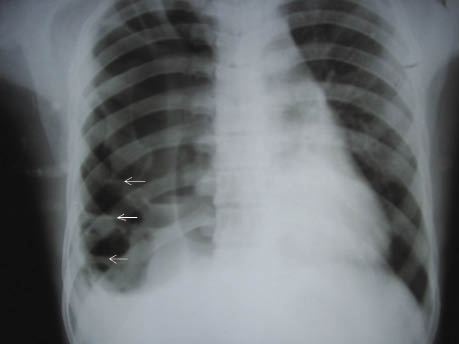
4. Pneumatocoeles: are intrapulmonary air-filled cystic spaces that can have a variety of sizes and appearances. Features are- smooth inner margins,contain little if any fluid, wall (if visible) is thin and regular, persist despite absence of symptoms.
Reading “Multiple Cystic translucent areas in the RLZ”
Round Pneumonia ( Rarely important) : Cause Bacterial infection in Children.
Round pneumonias are round-ish and while they are well-circumscribed parenchymal opacities, they tend to have irregular margins. Most commonly are solitary. Air-bronchograms are often present.

Next Article will include Chest Xray reading for Pulmonary TB, Collapse, Effusion and Abscess in next article.
If you have any comments, do send the feedback below.