
Resting Membrane Potential, Action Potential and Ionic Basis
Resting Membrane Potential (RMP)
There exists a potential difference across the cell membrane in all the living cells in resting conditions which is known as resting membrane potential. The value of RMP normally varies from 5 millivolts(mV) to -100 millivolts(mV). The value mainly depends upon the type of the cell. The value of RMP in nerve fibres and skeletal muscles is -90mV.
How is Resting Membrane Potential generated?
There are mainly four factors responsible for the genesis of Resting Membrane Potential. These are:
- The difference between the intracellular and extracellular potassium(K+) ion concentration.
- The impermeable nature of membrane to protein ions.
- Poor permeability of membrane to sodium(Na+) ions.
- The provision of sodium-potassium (Na+/K+ ATPase) pump in the membrane.
Action Potential
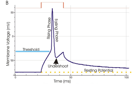
Action Potential is seen in all the excitable cells as well as some of the reproductive and endocrine cells. It basically means the transient change in membrane potential. Action potential is characterized by gradual depolarization to threshold to the rapid ascent followed by repolarization of the membrane. Action potential is comprised of the following stages:
- Resting Stage: It is the polarized stage of the membrane. The resting membrane potential (RMP) present in this stage is -90mV.
- Depolarization: In this stage the membrane suddenly becomes very permeable to sodium (Na+) ions. The initially present polarized state is neutralized by inflow of Na + ions. Then, the membrane potential rises rapidly in positive direction causing RMP to change from -90mV to -70mV, -50mV to 0mV and finally to +30mV.
- Repolarization: The repolarization of the membrane is characterized by the rapid diffusion of Potassium(K+) ions to exterior which re-establishes normal RMP. The diffusion of potassium ions takes place due to opening of potassium channels. Within (1/10000)th fraction of second after membrane becomes permeable to sodium(Na+) ions , the sodium ion channels begins to close and potassium ion channels open.
- Hyperpolarization: After passing through repolarization, there comes a stage when membrane potential becomes more negative than the resting membrane potential. The nerve at this stage is said to be hyperpolarized.
Ionic Basis of Action Potential:
There are various ionic key-players and ionic events responsible for the generation of “Action Potential” in the nerve fibre. The ionic basis of action potential in nerve fibre is discussed in the in following points:
- In resting state, cell membrane is more permeable to potassium (K + ) ions due to which very few sodium ion channels remain open.
- Depolarizing phase is caused due to opening of voltage gated sodium channels. This event increases sodium permeability by several hundred folds causing the potential difference across the membrane to become more positive.
- Repolarization phase occurs due to inactivation of voltage gated sodium ion channels and opening of voltage gated potassium channels which opens more slowly than sodium channels in response to depolarization.
- Some of the voltage gated potassium (K+) channels are still open which causes efflux of K + ions. This will give rise to more negative membrane potential than RMP which is called as hyperpolarization.
- Finally, after passing through all these stages, the re-establishment of resting membrane potential (RMP) takes place. Sodium-potassium pump plays major role in this restoration process.
Article by: Binaya Kafle
Facebook: http://facebook.com/brk.chekhob
Twitter: http://twitter.com/themedicalway
simplified and helpful.
Thank you